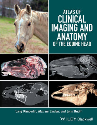
Stock image for illustration purposes only - book cover, edition or condition may vary.
Atlas of Clinical Imaging and Anatomy of the Equine Head
Larry Kimberlin
€ 164.68
FREE Delivery in Ireland
Description for Atlas of Clinical Imaging and Anatomy of the Equine Head
Hardcover. Atlas of Clinical Imaging and Anatomy of the Equine Head presents a clear and complete view of the complex anatomy of the equine head using cross-sectional imaging. Num Pages: 160 pages. BIC Classification: MZD; MZH. Category: (P) Professional & Vocational. Dimension: 222 x 285 x 13. Weight in Grams: 680.
Atlas of Clinical Imaging and Anatomy of the Equine Head presents a clear and complete view of the complex anatomy of the equine head using cross-sectional imaging. * Provides a comprehensive comparative atlas to structures of the equine head * Pairs gross anatomy with radiographs, CT, and MRI images * Presents an image-based reference for understanding anatomy and pathology * Covers radiography, computed tomography, and magnetic resonance imaging
Atlas of Clinical Imaging and Anatomy of the Equine Head presents a clear and complete view of the complex anatomy of the equine head using cross-sectional imaging. * Provides a comprehensive comparative atlas to structures of the equine head * Pairs gross anatomy with radiographs, CT, and MRI images * Presents an image-based reference for understanding anatomy and pathology * Covers radiography, computed tomography, and magnetic resonance imaging
Product Details
Publisher
John Wiley and Sons Ltd
Format
Hardback
Publication date
2016
Condition
New
Weight
680 g
Number of Pages
160
Place of Publication
Hoboken, United States
ISBN
9781118988978
SKU
V9781118988978
Shipping Time
Usually ships in 7 to 11 working days
Ref
99-11
About Larry Kimberlin
Larry Kimberlin, DVM, FAVD, CVPP, is the owner of Northeast Texas Veterinary Dental Center in Greenville, Texas, USA. Alex zur Linden, DVM, DACVR, is Assistant Professor of Radiology at Ontario Veterinary College at the University of Guelph in Ontario, Canada. Lynn Ruoff, DVM is Clinical Associate Professor at Texas A&M University ... Read more
Reviews for Atlas of Clinical Imaging and Anatomy of the Equine Head
Atlas of Clinical Imaging and Anatomy of the Equine Head is a comprehensive reference of the cross-sectional anatomy of the head of equids that features photographs of gross sections, CT images, and MRI scans of the head in transverse, sagittal, and dorsal planes. The photographs of gross-section preparations are excellent, and most anatomic features are readily identifiable. Furthermore, the anatomic ... Read more
